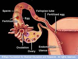
OVIDUCT - ITS TYPES & THEIR ROLE IN PREGNANCY
In the pregnant oviducts did not exceed 30 microns In the ovarian
end and 60 microns In the mid-reglon.
These can be classified into both
In the mature ducts the musculature showed a maximum measurement of 75 microns In the mid-reglon and 45 microns in the ovarian end. This showed that there were cyclic changes in the tunica muscularis of mature ducts, and that the musculature was limited to its minimum during pregnancy. As the growth cycle in the oviducts and the uterus of pregnant animals was not similar, it may also be presumed that the hormonal action which brought the changes in the two organs may be different and unrelated.

WHAT DO YOU MEAN BY IMMATURE OVIDUCTS ?
The immature oviducts, considered as a whole, resembled
closely the mature duct in structure but exhibited the following
differences:
1) The folds or plicae were fewer and simpler in their pattern.
2) The epithelium was simple, ciliated columnar without pseudostratification and contained a large number of spherical lymphocytes.
3) The epithelial cells were loosely ar- ranged and there was no cellular activity as would be indicated by protoplasmic protrusions or nuclear extrusions.
4) The elastic fibers in the tunica muscularis were less numerous than in the mature ducts.
5) Because the folds were few and simple.
The mature ducts were collected at different stages of the estrous cycle. The ducts from pregnant animals were
collected during the early part of the pregnancy suid immature
ducts were collected from calves varying from three weeks to nine
months of age.


Serial sections were made from each duct at three
different regions, namely the ovarian end, the mid-region and the
uterine end. The sections were stained by hematoxylin and eosin
and also special connective tissue stains. The three coats of the
duct, the tunica mucosa, muscularis and serosa were described in
detail.


The distinct characteristics of various regions of the
oviduct in regard to the number of mucosal folds, the pseudo-
stratified condition of the epithelium, the cellular activity
and the variations in the thickness of the muscular coats were
noted. The differences in structure exhibited by the ducts from
pregnant animals as compared to the mature ducts, which were con-
fined mostly to cytoplasmic protrusions, were reported.
No comments:
Post a Comment
Welcome to Bulky Uterus ... Post your Comments Here ....